DESK RELIEF Colchonete • Iniciante
Discs: L1-L2, L2-L3, L3-L4, L4-L5, L5-S1
Alongamento do Psoas em Ajoelhado
The psoas muscle attaches directly to the front of the lumbar vertebrae (L1-L5) and the femur. When shortened from sitting, it pulls the lower back into hyperlordosis and anterior pelvic tilt. Lengthening it removes immense lumbar shear stress.
DIAGRAMA BIOMECÂNICO DO MOVIMENTO
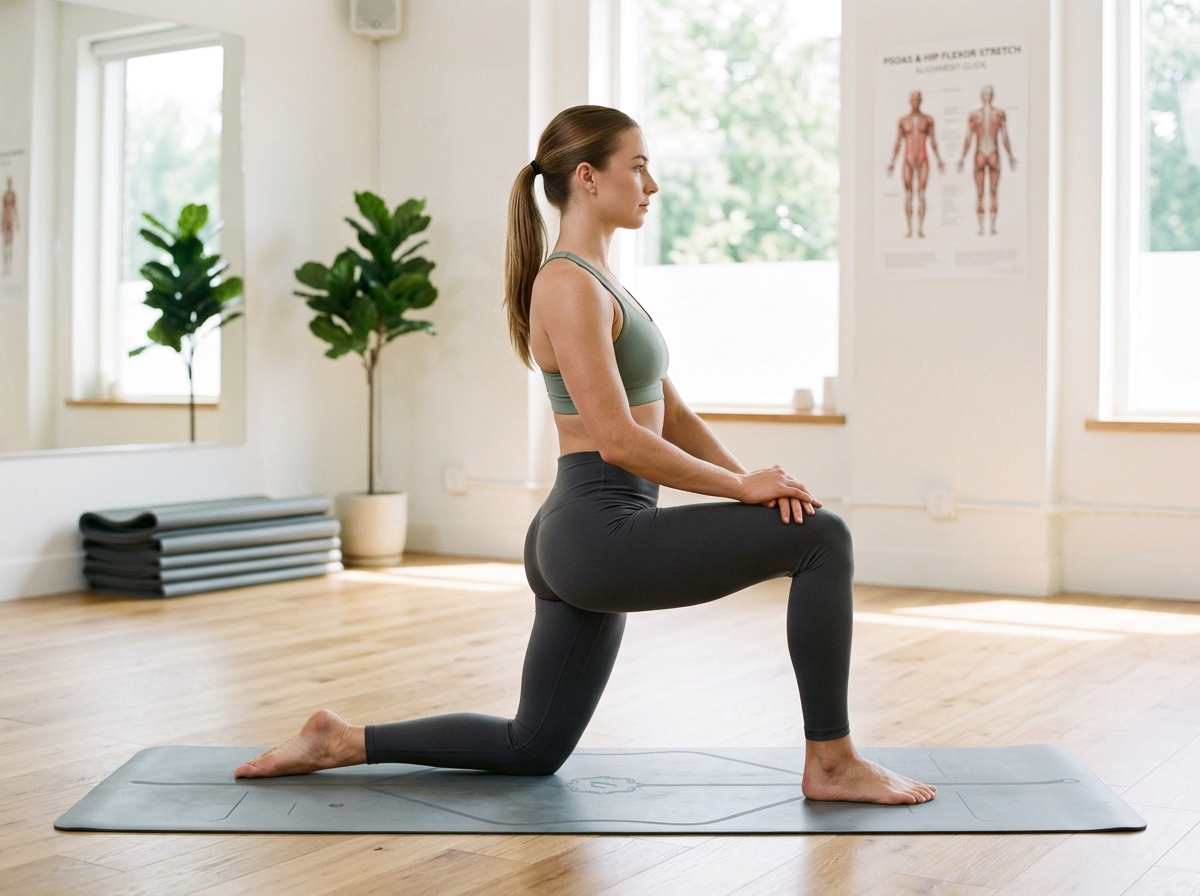
PSOAS & HIP DECOMPRESSION
🎯 Anterior Hip Flexors
Execução Passo a Passo
PROTOCOLO CLÍNICO- 1 Begin in a half-kneeling position with one knee on a padded mat and the front foot flat on the floor, both knees at 90 degrees.
- 2 Tuck your pelvis underneath you (posterior pelvic tilt) and squeeze the glute on the kneeling side.
- 3 Keeping your torso upright, gently shift your entire body weight forward by 1-2 inches until you feel a deep stretch in the front of your kneeling hip.
- 4 Optionally reach the arm on the kneeling side straight overhead and lean slightly toward the opposite side.
- 5 Hold for 30 seconds while breathing calmly. Switch sides and repeat.
🎯
PONTOS-CHAVE DE POSTURA
- ✓ Do not arch your lower back to lean forward; keep your pelvis tucked and spine tall.
- ✓ Squeeze the glute on the kneeling leg throughout the stretch to reciprocal-inhibit the hip flexor.
⚠️
ERROS A EVITAR
- ✗ Do not hyperextend your lumbar spine to get deeper forward.
- ✗ Avoid letting your front knee collapse inward.
🛑
CONTRAINDICAÇÕES E QUANDO EVITAR
Do not perform this movement or modify strictly under clinical supervision if you have:
- Severe knee joint pain or patellar bursitis on kneeling leg (use thick cushion or standing variation)
FUNDAMENTOS CIENTÍFICOS & CITAÇÕES
Biomecânica Clínica & Evidências
This exercise protocol utilizes proven spinal mechanics: active core co-contraction (McGill stabilization), directional preference extension (McKenzie MDT), and non-compressive axial elongation to facilitate nutrient exchange in the avascular intervertebral disc matrix.
• McGill, S.M. (2016). Low Back Disorders: Evidence-Based Prevention and Rehabilitation. Human Kinetics.
• Delitto, A., et al. (2021). Low Back Pain Clinical Practice Guidelines. Journal of Orthopaedic & Sports Physical Therapy (JOSPT).
DOSAGEM CLÍNICA
Tempo de Sustentação 30 Seconds
Repetições 3 holds each side
Séries Prescritas 1 set
Dificuldade Iniciante
Posição Colchonete
MÚSCULOS ALVO
Iliopsoas Rectus Femoris Tensor Fasciae Latae
MÚSCULOS ESTABILIZADORES
Gluteus Maximus (opposite) Abdominal Wall